Services Provided
During your initial visit to The Glaucoma Center, there are a series of diagnostic tests that will be performed along with your comprehensive eye exam. Our state-of-the-art equipment uses the most advanced technology to determine whether or not you have glaucoma and if so, how severe it may be. Our doctors will review testing and eye examination results with you, and go over treatment options if needed. After your initial consultation, these tests may be performed again on a yearly basis or as deemed necessary by our doctors, to ensure that every effort is made to preserve your vision.
Due to the time this testing may take, we ask that you please allow two hours for the initial glaucoma evaluation.
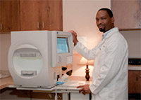
 Visual Field Testing:
This
is an automated examination of your peripheral (side) vision. One of
the complications of glaucoma can be loss of peripheral vision. This
test may show signs of damage before you become aware of it.
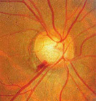
 Optic Disc Photography:
These are color photographs of the back of the eye, specifically the
optic nerve, which can be viewed in 3-dimensions by our doctors. Your
eye will need to be dilated for this type of photograph. It is important
to have these photographs so that your optic nerves can be followed
over time, and we can determine if any change has occurred by comparing
old photographs with your eye's current appearance.
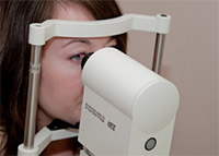
 Optical Coherence Tomography
These
are computerized images of the back of your eye. Your eye may or
may not require dilation to take these photographs. These tests measure
the size of the optic nerve, as well as the layer of retinal tissue
surrounding the optic nerve, the nerve fiber layer (NFL). This layer of
the retina can demonstrate early signs of glaucoma damage and can also
be monitored for any changes over time.
This imaging technique provides high-resolution, cross-sectional images of the retinal nerve fiber layer and the optic nerve head. This technology allows sensitive monitoring for glaucoma progression.